-
PROVIDERS
Learn more
Join Tempus at the 2026 ASCO® Annual Meeting!
-
LIFE SCIENCES
REGISTER NOW
From insight to impact: Leveraging the AI-enabled Next platform with BMS to advance equitable access in precision oncology
-
PATIENTS
It's About Time
View the Tempus vision.
- RESOURCES
-
ABOUT US
View Job Postings
We’re looking for people who can change the world.
- INVESTORS
AI-ENABLED PATHOLOGY
Our suite of digital pathology solutions use artificial intelligence to help you increase diagnostic confidence, improve workflow efficiency, and uncover deeper insights from every sample.
Contact usAssisted detection across diverse cancer types
-
PAIGE PROSTATE
A suite of AI-powered applications that aid in the detection and grading of prostate cancer on H&E-stained whole-slide images.
Paige Prostate Detect was the first AI-based software application in pathology to receive FDA authorization to aid in the primary diagnosis of prostate cancer.*
-
Paige Prostate Detect* | CE-IVDR, FDA-AUTHORIZED
AI application that helps pathologists find possible cancer in digital images of prostate biopsy slides. Paige Prostate Detect should be used in combination with specific scanners and viewers, and should only be used to support, not replace, a pathologist’s full evaluation.
Assists in the detection of even minute foci on slides that are suspicious for cancer. Offers high patient-level sensitivity and negative predictive value in cancer detection while decreasing pathologists’ reading times by 20%.1,2
-
Paige Prostate Grade & Quantify† | CE-IVDR, RUOCategorizes areas suspicious for cancer by Gleason pattern, providing predictions for primary and secondary Gleason grades. It also offers percentage and linear measurements of the tumor burden.
-
Paige Prostate Perineural Invasion (PNI)† | CE-IVDR, RUOAids in the detection of suspicious foci around nerve fibers to identify the presence of PNI, helping to streamline the final diagnosis.
* CE-IVDR for use with whole slide images visualized with the Paige FullFocus Image Viewer and digitized with Philips Ultra Fast Scanner, Leica Aperio AT2 Scanner, Leica GT450 Scanner, or Hamamatsu S360 Scanner. In the US, Paige Prostate Detect is FDA-authorized for clinical use. Paige Prostate Detect is a software only device intended to assist pathologists in the detection of foci that are suspicious for cancer during the review of scanned whole slide images (WSI) from prostate needle biopsies prepared from hematoxylin & eosin (H&E) stained formalinfixed paraffin embedded (FFPE) tissue. After initial diagnostic review of the WSI by the pathologist, if Paige Prostate Detect detects tissue morphology suspicious for cancer, it provides coordinates (X,Y) on a single location on the image with the highest likelihood of having cancer for further review by the pathologist. Paige Prostate Detect is intended to be used with slide images digitized with Philips Ultra Fast Scanner and visualized with the Paige FullFocus Image Viewer. Paige Prostate Detect is an adjunctive computer-assisted methodology and its output should not be used as the primary diagnosis. Pathologists should only use Paige Prostate Detect in conjunction with their complete standard of care evaluation of the slide image.
† CE-IVDR for use with whole slide images visualized with the Paige FullFocus Image Viewer and digitized with Philips Ultra Fast Scanner, Leica Aperio AT2 Scanner, Leica GT450 Scanner, or Hamamatsu S360 Scanner. For Research Use Only (RUO) in the US. Not for use in diagnostic procedures in the US.
-
-
PAIGE BREAST
A suite of AI-powered applications for research use only to support increasing efficiency in breast cancer detection and staging.
-
Paige Breast Detect & Neoplasm‡ | RUOHighlights areas most likely to harbor cancer and in situ neoplasms, focusing your review on critical regions of the slide.
-
Paige Breast Mitosis‡ | RUOIdentifies mitotic hotspots and provides a count and density of mitotic instances, allowing you to easily review individual mitotic figures to aid in grading.
-
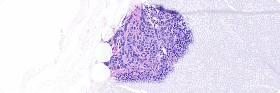
Paige Breast Lymph Node‡ | RUODetects foci suspicious for breast cancer in lymph node specimens. A study of anatomic pathologists demonstrated that use of Paige Breast Lymph Node improved pathologist sensitivity while cutting slide read times by over half.3
‡ For Research Use Only (RUO). Not for use in diagnostic procedures.
-
-
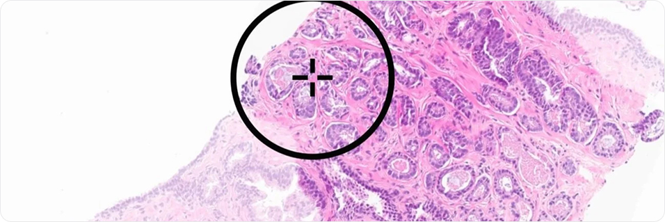
PAIGE PANCANCER
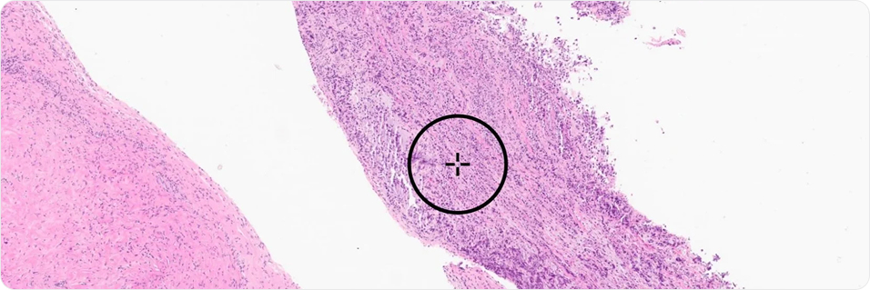
A groundbreaking application for research use only, capable of identifying foci suspicious for cancer across more than 40 cancer types, developed using Paige’s foundation model for computational pathology.
-
Paige PanCancer Detect‡ | RUODetect suspicious tissue across more than 40 cancer types to support case triage with prediagnostic screening of all biopsy and resected tissue, or postdiagnostic QC. Achieved 0.95 specimen-level AUC across nine common and seven rare cancers.4
‡ For Research Use Only (RUO). Not for use in diagnostic procedures.
-
-
LIMITED FEEDBACK RELEASE
We continue to expand our suites of digital pathology solutions for research. Additional AI applications are currently available on a limited basis for research use only feedback.
- Paige Breast Detect & Subtype‡ | RUO
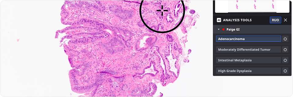
- Paige GI Detect & Subtype‡ | RUO
- Paige GYN Detect & Subtype
- Paige Bladder Detect & Subtype
‡ For Research Use Only (RUO). Not for use in diagnostic procedures.
A suite of AI-powered applications that aid in the detection and grading of prostate cancer on H&E-stained whole-slide images.
Paige Prostate Detect was the first AI-based software application in pathology to receive FDA authorization to aid in the primary diagnosis of prostate cancer.*
-
Paige Prostate Detect* | CE-IVDR, FDA-AUTHORIZED
AI application that helps pathologists find possible cancer in digital images of prostate biopsy slides. Paige Prostate Detect should be used in combination with specific scanners and viewers, and should only be used to support, not replace, a pathologist’s full evaluation.
Assists in the detection of even minute foci on slides that are suspicious for cancer. Offers high patient-level sensitivity and negative predictive value in cancer detection while decreasing pathologists’ reading times by 20%.1,2
-
Paige Prostate Grade & Quantify† | CE-IVDR, RUOCategorizes areas suspicious for cancer by Gleason pattern, providing predictions for primary and secondary Gleason grades. It also offers percentage and linear measurements of the tumor burden.
-
Paige Prostate Perineural Invasion (PNI)† | CE-IVDR, RUOAids in the detection of suspicious foci around nerve fibers to identify the presence of PNI, helping to streamline the final diagnosis.
* CE-IVDR for use with whole slide images visualized with the Paige FullFocus Image Viewer and digitized with Philips Ultra Fast Scanner, Leica Aperio AT2 Scanner, Leica GT450 Scanner, or Hamamatsu S360 Scanner. In the US, Paige Prostate Detect is FDA-authorized for clinical use. Paige Prostate Detect is a software only device intended to assist pathologists in the detection of foci that are suspicious for cancer during the review of scanned whole slide images (WSI) from prostate needle biopsies prepared from hematoxylin & eosin (H&E) stained formalinfixed paraffin embedded (FFPE) tissue. After initial diagnostic review of the WSI by the pathologist, if Paige Prostate Detect detects tissue morphology suspicious for cancer, it provides coordinates (X,Y) on a single location on the image with the highest likelihood of having cancer for further review by the pathologist. Paige Prostate Detect is intended to be used with slide images digitized with Philips Ultra Fast Scanner and visualized with the Paige FullFocus Image Viewer. Paige Prostate Detect is an adjunctive computer-assisted methodology and its output should not be used as the primary diagnosis. Pathologists should only use Paige Prostate Detect in conjunction with their complete standard of care evaluation of the slide image.
† CE-IVDR for use with whole slide images visualized with the Paige FullFocus Image Viewer and digitized with Philips Ultra Fast Scanner, Leica Aperio AT2 Scanner, Leica GT450 Scanner, or Hamamatsu S360 Scanner. For Research Use Only (RUO) in the US. Not for use in diagnostic procedures in the US.
A suite of AI-powered applications for research use only to support increasing efficiency in breast cancer detection and staging.
-
Paige Breast Detect & Neoplasm‡ | RUOHighlights areas most likely to harbor cancer and in situ neoplasms, focusing your review on critical regions of the slide.
-
Paige Breast Mitosis‡ | RUOIdentifies mitotic hotspots and provides a count and density of mitotic instances, allowing you to easily review individual mitotic figures to aid in grading.
-
Paige Breast Lymph Node‡ | RUODetects foci suspicious for breast cancer in lymph node specimens. A study of anatomic pathologists demonstrated that use of Paige Breast Lymph Node improved pathologist sensitivity while cutting slide read times by over half.3
‡ For Research Use Only (RUO). Not for use in diagnostic procedures.
A groundbreaking application for research use only, capable of identifying foci suspicious for cancer across more than 40 cancer types, developed using Paige’s foundation model for computational pathology.
-
Paige PanCancer Detect‡ | RUODetect suspicious tissue across more than 40 cancer types to support case triage with prediagnostic screening of all biopsy and resected tissue, or postdiagnostic QC. Achieved 0.95 specimen-level AUC across nine common and seven rare cancers.4
‡ For Research Use Only (RUO). Not for use in diagnostic procedures.
We continue to expand our suites of digital pathology solutions for research. Additional AI applications are currently available on a limited basis for research use only feedback.
- Paige Breast Detect & Subtype‡ | RUO
- Paige GI Detect & Subtype‡ | RUO
- Paige GYN Detect & Subtype
- Paige Bladder Detect & Subtype
‡ For Research Use Only (RUO). Not for use in diagnostic procedures.
Tissue can be scarce, insights don’t have to be
The Paige Predict suite of applications help optimize tissue stewardship and inform testing strategies before exhausting tissue.
Paige Predict: Tissue Optimization | RUO
AI application to help inform research decisions prior to molecular profiling
Forecasts total nucleic acid (TNA) yield from an H&E image to identify samples at risk of NGS failure.
Paige Predict: Biomarker Prediction | RUO
AI application predicting the status of ~1,600 biomarkers across 505 genes from a single H&E slide.
Accelerate biomarker screening across samples
Provides results in approximately 5 minutes to accelerate sample selection for testing across NGS, hotspot testing, IHC, and special stains.
Receive genomic insights across cancer types
Predictions can help researchers prioritize use of tissue, especially in cancers with often limited tissue (e.g., NSCLC) or less well tested indications (e.g., Melanoma, Endometrium).
Enrich cohorts for research at scale
Pre-screen samples for biomarkers of interest to help enable relevant sample identification at scale, supporting research and time to enrollment in clinical studies.
Paige Predict features are for Research Use Only (RUO) and are not for use in diagnostic procedures. Paige Predict results are probabilistic predictions and should not be used to determine treatment eligibility or as evidence of actual biomarker status.
Paige Predict deployed at Tempus
Tempus has deployed Paige Predict applications into our standard NGS lab workflow, with no additional ordering steps required.
Tissue optimization
Tempus has integrated Paige Predict’s tissue optimization capabilities into our clinical laboratory workflow to help our pathologists conserve tissue and maximize the chances of a successful result. This process has reduced tissue utilization by 18% and is expected to lower the overall QNS rate by 15%.5
Biomarker prediction
Paige Predict’s biomarker predictions for 123 clinically relevant features across 16 cancer cohorts have been validated for clinical use as a Tempus laboratory developed test and is performed for eligible xT QNS cases, helping to inform confirmatory testing strategies.
Paige Predict results are probabilistic predictions and should not be used to determine treatment eligibility or as evidence of actual biomarker status.
Flexible deployment for your laboratory
Choose the deployment model that fits your laboratory’s research needs, with options for integration with your existing platform or our image management system (IMS).
- Access Paige applications within your current digital pathology ecosystem, maintaining use of your preferred scanner, IMS, and viewer.
- Use the Paige IMS, a secure, cloud-native platform that includes the FullFocus® Image Viewer*, FullFolio™ case management, and an application marketplace for third-party AI.
*In the United States, FullFocus® is cleared for clinical use with Philips Ultrafast Scanner, Leica Aperio GT 450 DX, and Hamamatsu NanoZoomer S360MD Slide scanner system. FullFocus version 2.29 was not validated for use with images generated with Philips Ultra Fast Scanner.
Paige IMS
Workflow Integration
-
AI-Native Web Viewer
FullFocus® provides high-performance visualization accessible from any authorized location.
-
Hardware Agnostic
Seamlessly integrates with existing digital scanners, including Philips, Leica, and Hamamatsu.
-
Cloud-Native Scale
Flexible case management (FullFolio™) and storage designed for large-scale digital pathology without the need for on-premise hardware.
Our Science
view all publications-
UPCOMING WEBINAR:
Independent real-world application of a clinical-grade automated prostate cancer detection system
Read publication -
UPCOMING WEBINAR:
Clinical Validation of Artificial Intelligence–Augmented Pathology Diagnosis Demonstrates Significant Gains in Diagnostic Accuracy in Prostate Cancer Detection
Read publication -
UPCOMING WEBINAR:
Artificial Intelligence Helps Pathologists Increase Diagnostic Accuracy and Efficiency in the Detection of Breast Cancer Lymph Node Metastases
Read publication -
UPCOMING WEBINAR:
A foundation model for clinical-grade computational pathology and rare cancers detection
Read publication
- da Silva, LM, Pereira, EM, Salles PG, et al. Independent real‐world application of a clinical‐grade automated prostate cancer detection system. J Pathol. 2021;254(2):147-158.
- Eloy, Catarina, et al. Artificial intelligence–assisted cancer diagnosis improves the efficiency of pathologists in prostatic biopsies. Virchows Archiv. 482.3 (2023): 595-604.
- Retamero Juan Antonio, Emre Gulturk, Bozkurt A, et al. Artificial intelligence helps pathologists increase diagnostic accuracy and efficiency in the detection of breast cancer lymph node metastases. Am J Surg Pathol. 48.7 (2024): 846-854.
- Vorontsov E, Bozkurt A, Casson A, et al. A foundation model for clinical-grade computational pathology and rare cancers detection. Nat Med 30, 2924–2935 (2024).
- Tempus AI, Inc. Data on file. 2025.
Bring AI-enabled pathology to your laboratory
By combining Paige’s industry-leading digital pathology expertise with the broader Tempus ecosystem, we are redefining what’s possible in AI-enabled pathology to empower anatomic and molecular pathologists and researchers worldwide.