-
PROVIDERS
Register now
Are you getting the full picture? A webinar series on the power of comprehensive intelligent diagnostics
-
LIFE SCIENCES
Enroll now
Tempus’ Patient-Derived Organoid ScreensEvaluate the efficacy of your preclinical compounds using fixed organoid panels designed for diverse therapeutic applications. Space is limited — enroll by June 30, 2025, to secure your spot.
-
PATIENTS
It's About Time
View the Tempus vision.
- RESOURCES
-
ABOUT US
View Job Postings
We’re looking for people who can change the world.
- INVESTORS
RADIOLOGY /// LUNG
AI-enabled solution to detect and track longitudinal changes in lung nodules

Tempus Pixel offers advanced analysis and automated reporting from routine CT images to help improve efficiency and accuracy in detecting and tracking changes in lung nodules over time, aiding providers in making informed diagnostic and disease management decisions.
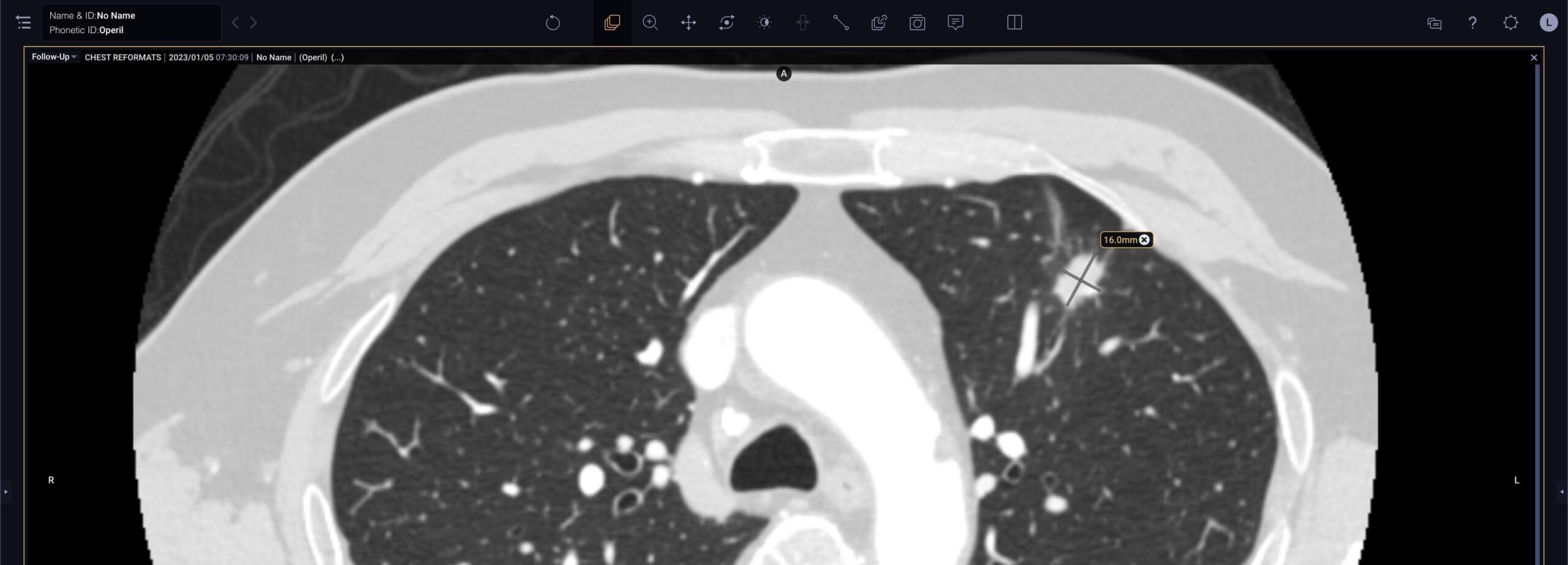
nodule detection
Automatically detects and segments lung nodules in CT images, helping providers to efficiently and accurately identify potentially significant findings1,2,3
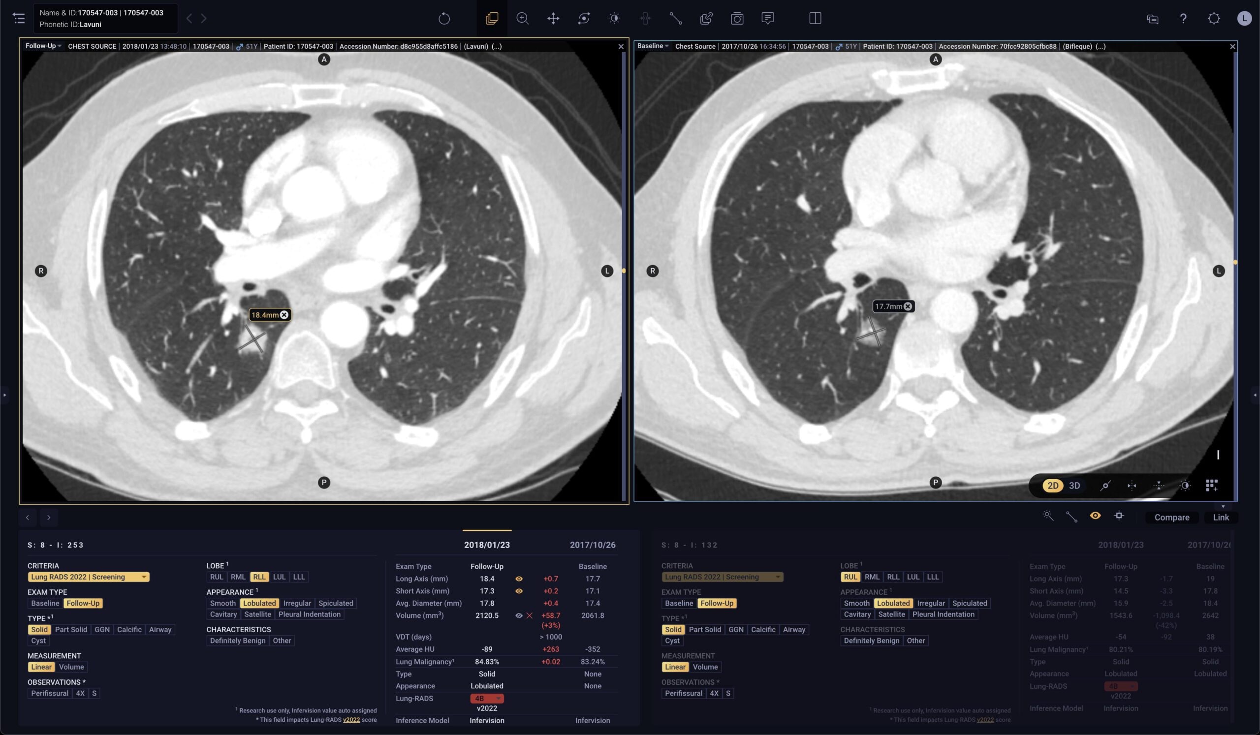
nodule quantification
Automatically quantifies and localizes lung nodules in Lung CT images to help providers make efficient, consistent, and accurate assessments1,2

nodule tracking
Automatically tracks changes in lung nodules across exams over time with tabular and graphical results for deeper insights into disease progression1,2
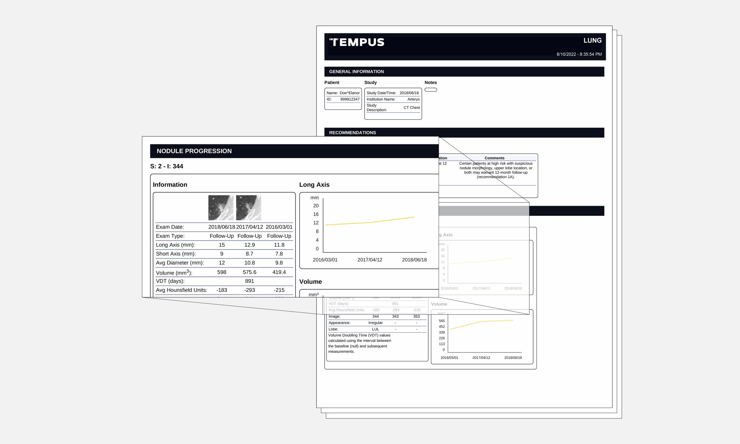
nodule reporting
Automatically builds standardized reporting that summarizes findings and relevant information, including scoring recommendations, for easy interpretation

Malignancy risk stratification
Improves the non-invasive stratification of CT identified nodules at the time of nodule detection with the nodule malignancy score. This feature is available for use in research applications4

Technical requirements
- Patients 18 years and older only
- Axial CT images only
- Images must be 3.2mm slice thickness or less
- Images must be sequential
- Data slice thickness requires slice gap ≤ slice thickness
- Images must include entire lung in FoV (e.g. abdominal studies not acceptable)
- DICOM header must include all the following tags: image position, patient location, slice location, slice thickness
- In the U.S., Tempus Pixel Lung is FDA-cleared (K203744) but is not indicated for lung nodule detection. Lung nodule automated detection and quantification is powered by InferRead CT (K192880). Arterys Inc is the manufacturer of Tempus Pixel Lung, excluding any third party components described in this list. Infervision is the manufacturer of InferRead CT.
- In the EU Tempus Pixel Lung is CE marked and indicated for lung nodule detection.
- Kozuka T, Matsukubo Y, Kadoba T, et al. Efficiency of a computer-aided diagnosis (CAD) system with deep learning in detection of pulmonary nodules on 1-mm-thick images of computed tomography. Jpn J Radiol. 2020;38(11):1052-1061. https://doi.org/10.1007/s11604-020-01009-0
- RUO indication for US and EU.