-
PROVIDERS
Register now
Are you getting the full picture? A webinar series on the power of comprehensive intelligent diagnostics
-
LIFE SCIENCES
Enroll now
Tempus’ Patient-Derived Organoid ScreensEvaluate the efficacy of your preclinical compounds using fixed organoid panels designed for diverse therapeutic applications. Space is limited — enroll by June 30, 2025, to secure your spot.
-
PATIENTS
It's About Time
View the Tempus vision.
- RESOURCES
-
ABOUT US
View Job Postings
We’re looking for people who can change the world.
- INVESTORS
11/08/2022
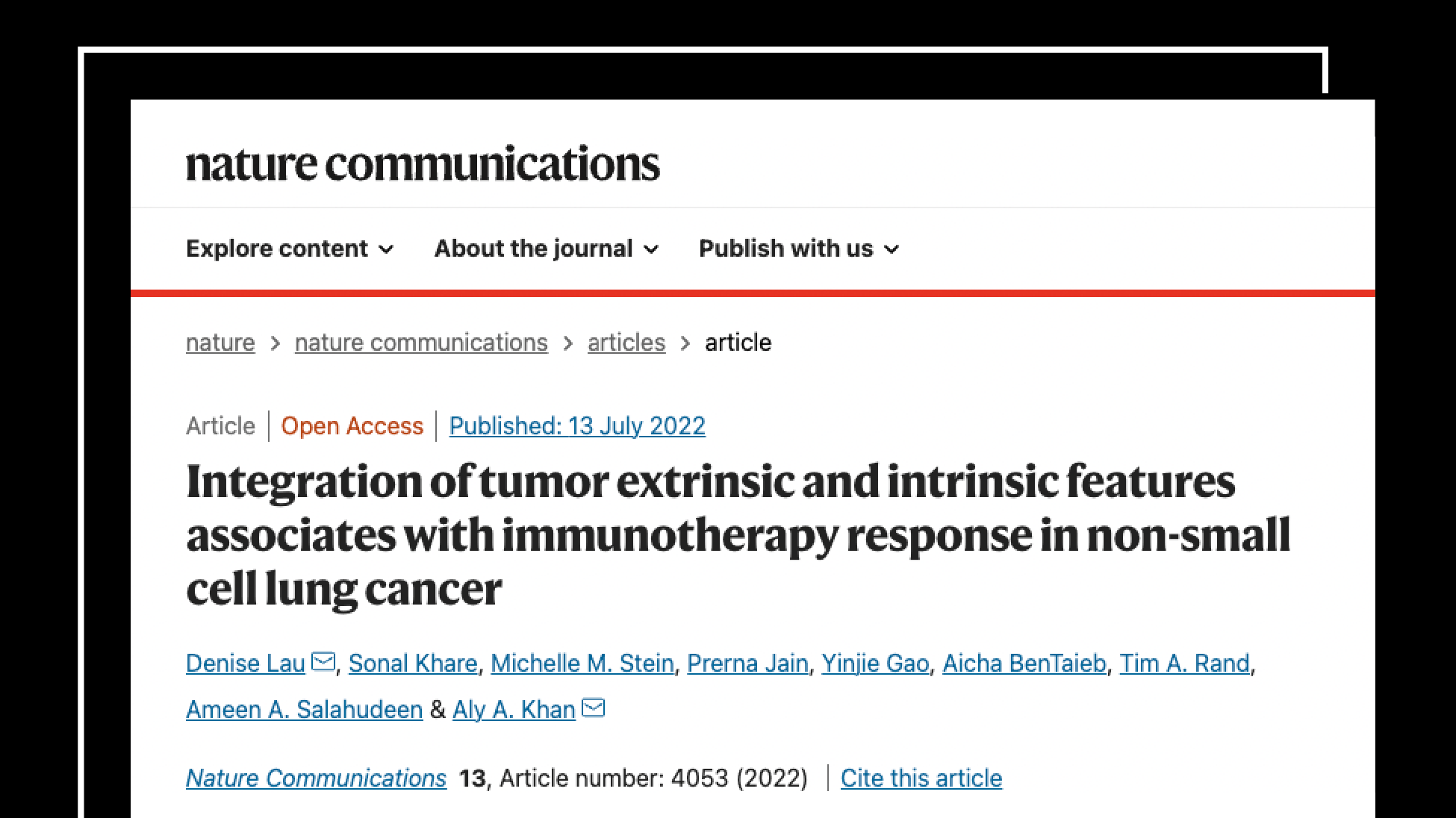
New research seeks to explain difference in ICB therapy response
Tempus developed an integrative model of tumor-extrinsic and tumor-intrinsic features associated with longer time to progression in NSCLC patients.
Authors
Michelle Stein, PhD
Director, Computational Biology, Tempus

Director, Computational Biology, Tempus

Research summary
In metastatic non-small cell lung cancer (mNSCLC), response rates to PD-(L)1 immune checkpoint blockade (ICB) therapies vary widely, and the mechanisms of response and resistance are not well understood. Previous studies have proposed disruption of HLA class I antigen presentation as an important mechanism of immune escape and ICB resistance, as this may interfere with direct killing of tumor cells by cytotoxic CD8+ T cells.1-3
With rapid advances in DNA and RNA sequencing, the use of linked clinical, genomics, and transcriptomics datasets, like those found in the Tempus Multimodal Database, can be used to facilitate analysis of transcriptional and genomic signatures associated with ICB response, supplementing evidence generated in clinical trials or other studies.
My coauthors and I sought to identify HLA-I-independent features that were associated with ICB response in mNSCLC. We turned to Tempus’s biological modeling laboratory to generate single-cell multiomic profiling datasets from NSCLC tumor specimens – scRNAseq, T cell receptor sequencing, and surface protein profiling – to characterize the tumor and T cell compartments in NSCLC tumors from 10 patients. We discovered a novel population of tumor-infiltrating, clonally expanded CD4+ helper T cells aberrantly expressing genetic programming similar to classical cytotoxic CD8+ killer T cells. In addition, tumor cells from these lung cancers also showed expression of the HLA class II, allowing them to be susceptible to these cytotoxic CD4+ T cells. These findings add to the emerging evidence (Oh et al., Cohen et al., Awad et al.) that cytotoxic CD4+ T cells are a noteworthy component of the tumor immune microenvironment and may play a role in anti-tumor immune responses following treatment with ICB.
We leveraged the single-cell multiomic data to develop a specific transcriptomic signature that captured both CD4+ and CD8+ cytotoxic T cells populations in tumors and applied it to a real-world cohort of mNSCLC patients selected from the Tempus Clinico-genomic Database (n=123). By combining this gene signature for cytotoxicity with tumor mutational burden (TMB), we developed an integrative model of tumor-extrinsic (cytotoxic gene signature) and tumor-intrinsic TMB features that is associated with longer time to progression in a real-world cohort of mNSCLC patients treated with ICB regimens, including those with disrupted class I HLA. These results demonstrate that integrating tumor-extrinsic and tumor-intrinsic features may be an informative biomarker for identifying mNSCLC patients who are more likely to respond to ICB and can remain effective even in populations where tumor HLA-LOH is common.
We are now working with our biopharma partners to apply this novel signature into their programs to improve patient selection. For example, biopharma is:
- Collaborating with Tempus to design and rapidly deploy novel single-cell experiments and compare real-world multimodal data aimed at translating ICB insights into clinical biomarkers.
- Applying our model of ICB response to enhance existing biomarkers of ICB therapies in both the metastatic and neoadjuvant setting in NSCLC and other cancers.
- Further exploring biomarkers of response or resistance to ICB or other therapies of interest in the Tempus Multimodal Database.
- Testing and further developing existing preclinical transcriptional signatures into actionable biomarkers.
Next steps
Contact Tempus to discuss this principled approach with one of our computational biologists. We can dive even deeper with you as it relates to results for your immunotherapy portfolio or design bespoke projects to advance your research and clinical programs.
References
- Sade-Feldman, M. et al. Resistance to checkpoint blockade therapy through inactivation of antigen presentation. Nat. Commun. 8, 1–11 (2017).
- Gettinger, S. et al. Impaired HLA class I antigen processing and presentation as a mechanism of acquired resistance to immune checkpoint inhibitors in lung cancer. Cancer Discov. 7, 1420–1435 (2017).
- Zaretsky, J. M. et al. Mutations associated with acquired resistance to PD-1 blockade in melanoma. N. Engl. J. Med. 375, 819–829 (2016).
READ THE MANUSCRIPT
Lau, D., Khare, S., Stein, M.M. et al. Integration of tumor extrinsic and intrinsic features associates with immunotherapy response in non-small cell lung cancer. Nat Commun 13, 4053 (2022). https://doi.org/10.1038/s41467-022-31769-4
-
04/02/2025
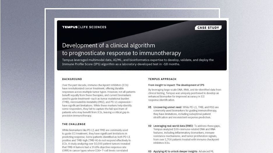
Development of a clinical algorithm to prognosticate response to immunotherapy
Discover how Tempus developed and deployed the Immune Profile Score (IPS)—a powerful algorithm that provides prognostic insights into patient outcomes following treatment with immune checkpoint inhibitors (ICIs)—in ~18 months. This case study highlights the AI-driven methodology, real-world validation, and the impact of IPS in precision oncology.
Read more -
03/25/2025
AI & ML in action: Unlocking RWD with GenAI through Tempus Lens
Discover how Tempus is equipping researchers with innovative AI solutions to fully leverage the potential of multimodal data. Gain insights from a panel of leaders across healthcare and life sciences as they discuss the impact of these advanced tools on delivering insights with speed.
Watch replay
Secure your recording now. -
03/11/2025
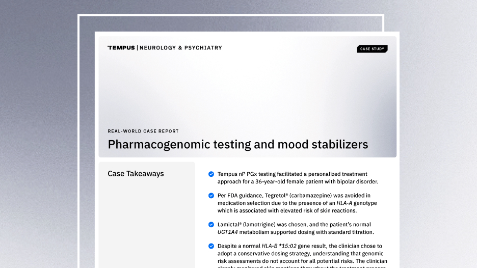
Case Report: Pharmacogenomic testing and mood stabilizers
This real-world case demonstrates how the Tempus nP pharmacogenomic test facilitated a personalized treatment approach for a patient with bipolar disorder.
Read more


